High Quality Rechargeable LED DE-3100 Dermatoscope

$499.00

$499.00
  • In Stock

  • Arrive in 5-7 days

  • Free Shipping Worldwide $59+

  • 2 Years Warranty

Info
Features & Compatibility
  • 10 x Magnification
  • 32mm wide field of view (Aperture)
  • Polarized, non-polarized, and amber lightillumination
  • Detachable protective glass
  • Automatic shutdown
  • Included adapter fits all phone
  • Bright LED illumination lights
  • All-metal housing
Specs
Material Aluminum & Glass
Optical Design All glass, 4 elements 3 groups, anti-reflection coating
Lens Diameter 32mm(front); 25mm(rear)
Magnification 10x
Distortion 8%
Polarization Cross Polarization
Resolution 300 LP/MM (Axis) 280 LP/MM (Edges)
LED 30 LEDs, 18 polarization, 6 non polarization, 6 amber polarization
Battery Capacity 1000mAh Lithium ion
Charging USB-C
Working Time 6-8 hours
Focus Distance 12-20mm
Dimension Φ53mm*H35mm*L136.5mm
Weight 187g


$499.00

$499.00
  • In Stock

  • Arrive in 5-7 days

  • Free Shipping Worldwide $59+

  • 2 Years Warranty

How to Use

Check out our step-by step quick start guide of the device.

Play Video

What Makes it Unique

It’s a handheld dermatoscope, but it contains a universal phone adapter meaning convenient to connect with any smartphone or tablet to capture images. Build with 4K resolution optics,polarized light, 10X magnification and 25mm wide field of view — the DE-3100 is perhaps the most super value dermatoscope we’ve ever seen. The visual filed is smaller(22%) than DE-4100, but the weight is 40% lighter.

Why Choose IBOOLO DE-3100 Dermatoscope

Sharp & Precise

Made from premium multi-coated and multi-element optics, the imaging performance is leading in the market. Optimized for the latest iPhone, rank only next to DE-4100.

Naked eye detection. 25mm visual field of view.

Sharp & Precise

Cross Polarization

In polarization mode, the polarization filter becomes activated and absorbs the surface light reflection (no immersion fluid required). It allows you to examine the colours, shapes and texture features of the skin lesion more clearly, more precise and more detailed.

Cross Polarization

Efficient Lighting

Built with polarized & non-polarized light, the DE-3100 offers two levels of color spectrum control to enhance imaging of deeper pigmentation.

Efficient Lighting

Easy to Use

The magnet attachment gives the ability to attach on smartphone and take pictures in 5 seconds.

Best part? It compatible with all phone in the market, with a universal lens clip or a phone cover for easy on and off.

Focus adjustable. It’s able to adjust working distance of different phone cameras. This enables the best image capture effect to be achieved.

Easy to Use

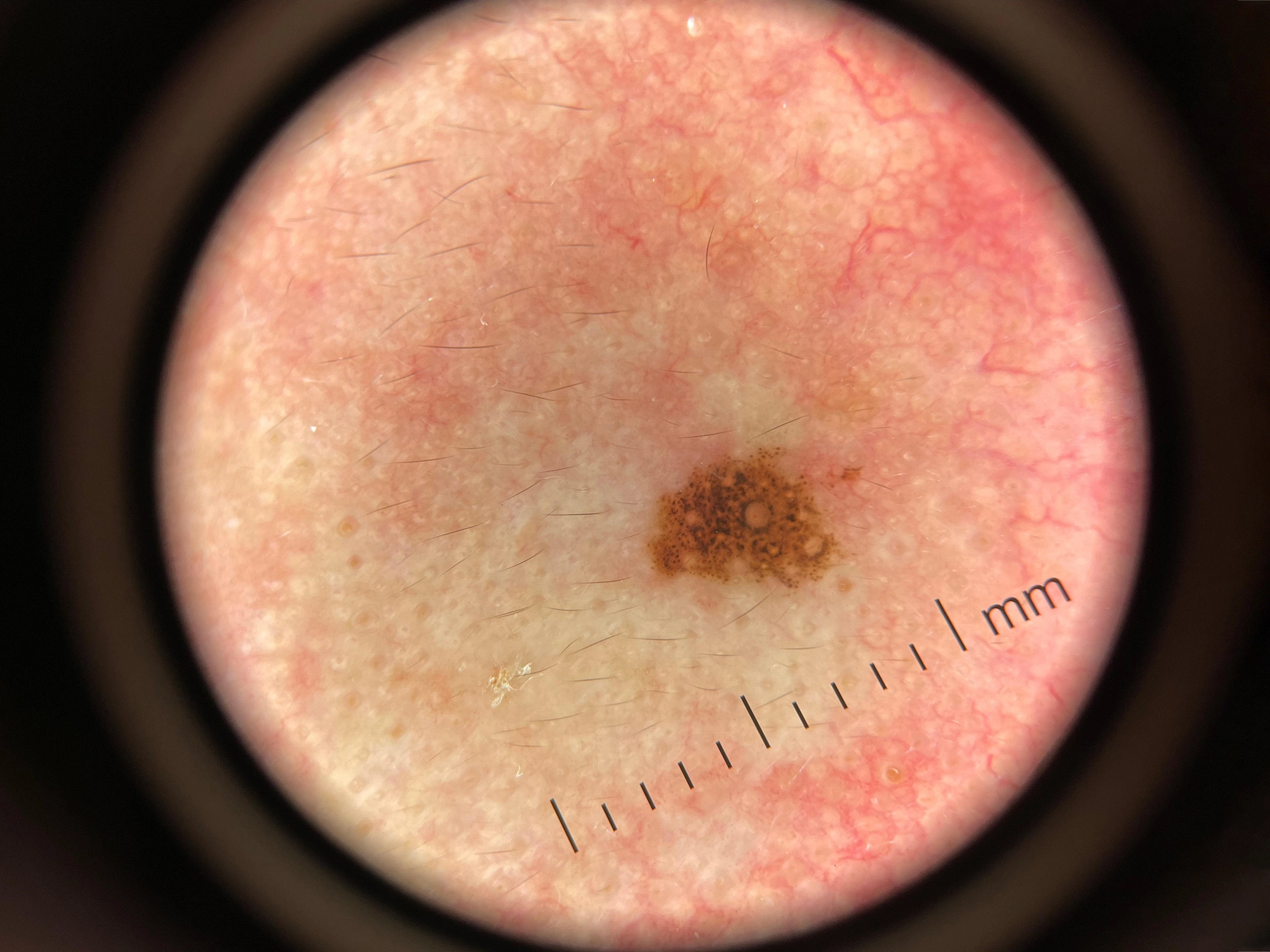
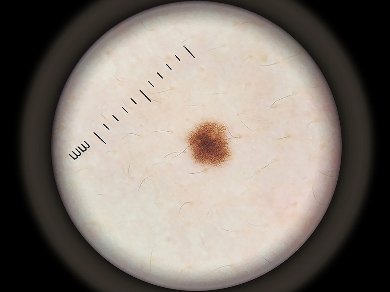
A Picture Speaks a Thousand Words

Toggle to hold the "after" image
DE-3100 Polarized EffectDE-3100 Non Polarized Effect

How Does it Compare

There’s an influx of dermatoscope on the market. Our devices are remarkable blend of pro-level features and affordable price. Their premium optics and efficient LED system delivers sharp & precise images.

  • OPTICAL DESIGN
  • LENS DIAMETER
  • MAGNIFICATION
  • LIGHTING SYSTERM
  • BATTERY
  • CONNECTABILITY
  • ADJUSTABLE
  • COST
output-onlinegiftools
Product LENS DIAMETER OPTICAL DESIGN MAGNIFICATION LIGHTING SYSTERM BATTERY CONNECTABILITY ADJUSTABLE COST

DE-4100

011
48mm/32mm 4 elements 3 groups 10 times 22 LEDs, polarise, non polarise, amber light 1000mAh naked eye, phone, tablet, digital camera focus & brightness $699.00

DE-3100

32mm/25mm 4 elements 3 groups 10 times 30 LEDs, polarise, non polarise, amber light 1000mAh naked eye, phone, tablet, digital camera focus $499.00

DE-500

de500
25mm/17mm 4 elements 3 groups 10 times 16 LEDs, polarise, non polarise, UV light 200mAh smartphone & tablet brightness $399.00

DE-400

DE-400 dermoscpy (01)
46mm/17mm 4 elements 3 groups 10 times 12 LEDs, polarization, non polarization 200mAh smartphone & tablet non adjustable $179.00

What’s in the Box?

DE-3100
  • DE-3100 Dermatoscope
  • Magnetic Phone Adapter
  • Charging Cable
  • Clean Cloth
  • Carrying Case
  • User Manual

Reviews

1 review for DE-3100 Dermatoscope

  1. Alan RosenbachCanada

    This dermatoscope is excellent. The attachment to the phone is a home run. Viewing is much easier. Great company with great communication

Only logged in customers who have purchased this product may leave a review.

Learn More

Skin Conditions in the Summer Cover

What Skin Conditions Are Most Common in the Summer?

Summer brings heat, humidity, strong sunlight, and increased outdoor activity. These environmental changes contribute to a rise in certain skin conditions. Among the most common are Miliaria, Polymorphous light eruption…

IBOOLO woods lamp cover

What Can a Wood’s Lamp Be Used For

A Wood’s lamp is a diagnostic device that emits long-wave ultraviolet (UV) light, typically in the range of 320 to 450 nm, with a peak emission around 365nm. When this…

How to Differentiate Rosacea, Acne, and Eczema cover

How to Differentiate Rosacea, Acne, and Eczema?

The skin, as the body’s largest organ, is susceptible to a myriad of inflammatory conditions. Among the most common are rosacea, acne, and eczema, which frequently present with overlapping symptoms…

0

No products in the cart.

Ask

Have questions on gear or your order?

Our Gear Guides are here to help! Get personal advice from pro creatives

Name
Subject
Email address
How can we help?
Answers

Instant Answers

How can dermoscopy images be captured?

Dermoscopy images can be captured and stored in different ways, such as: • Using a smartphone or tablet with dermoscopic adapter, which consisted in the package.• Using a digital camera

Dermoscopy images can be captured and stored in different ways, such as:

• Using a smartphone or tablet with dermoscopic adapter, which consisted in the package.
• Using a digital camera with dermoscopic adapter, there’s 49mm screw size camera adapter available to order now.

Compatible phone/tablet models:
All iPhone models, 95% Android phones, 90% tablet. For phone/tablet size in 5.25-14mm

Compatible camera models:
All camera with built 49mm filter screw, such as Canon EOS 70D, 80D, 90D; Canon EOS R7, R10, R50, R100; Canon M100, M200, M50, Mark II; Canon G7X Mark III, Sony ZV-1

How can I connect my phone to my dermatoscope?

There’s universal phone adapter for all our dermoscopes. Please check the installation procedure bellow or watch operation guide. Smartphone Connector (1) Place phone adapter screw in the center of smartphone’s

There’s universal phone adapter for all our dermoscopes. Please check the installation procedure bellow or watch operation guide.

Smartphone Connector

(1) Place phone adapter screw in the center of smartphone’s main camera.
(2) Screw magnet attachment on phone adapter.
(3) Put dermoscope’s back ring and magnet attachment together

Take The Best Images

You need to adjust the focus ring after the dermoscpe connected on smartphone to get the best images.

How can I clean my dermoscopy after usage?

Cleaning your dermoscopy after usage is important to prevent cross-contamination and infection. The cleaning method may vary depending on the type and model of your dermoscopy, so you should always

Cleaning your dermoscopy after usage is important to prevent cross-contamination and infection. The cleaning method may vary depending on the type and model of your dermoscopy, so you should always follow the manufacturer’s instructions. However, some general steps are:

• Turn off and disconnect your dermoscopy from any power source or device.

• Wipe off any visible dirt or debris from the dermoscopy with a soft cloth or tissue.

• Disinfect the dermoscopy with an alcohol-based wipe or spray, or a disinfectant solution recommended by the manufacturer. Make sure to cover all surfaces, especially the lens and contact plate.

• Let the dermoscopy air dry completely before storing it in a clean and dry place.

• Do not use abrasive or corrosive cleaners, solvents, or detergents that may damage the dermoscopy.

• Do not immerse the dermoscopy in water or any liquid, unless it is waterproof and designed for immersion.

You should clean your dermoscopy after each use, or at least once a day if you use it frequently. You should also check your dermoscopy regularly for any signs of damage or malfunction, and contact the manufacturer or service provider if needed.

Polarized VS Non-polarized Dermoscopy

A dermoscopy is a device that allows the examination of skin lesions with magnificationand illumination. By revealing subsurface structures and patterns that are not visible tothe naked eye. It can

A dermoscopy is a device that allows the examination of skin lesions with magnificationand illumination. By revealing subsurface structures and patterns that are not visible tothe naked eye. It can improve the diagnose accuracy of skin lesions, such as melanoma,basal cell carcinoma, seborrheic keratosis, etc.

There are two main types of dermoscopy: Non polarized and polarized dermoscopy.We’ve fitted most of our dermoscopys with polarized and non-polarized light. They canbe used in multiple skin structures.

Non-polarized contact Mode

In non-polarized mode, the instrument can provide information about the superficialskin structures, such as milia-like cysts, comedo-like openings, and pigment in theepidemis.

The dermoscopy requires applying a liquid such as mineral oil or alcohol to the skin andplacing the lens in contact with the skin. This reduces surface reflection and enhancesthe view of subsurface structures.

Image with non-polarized light (DE-3100)

Polarized contact Mode

In polarized mode, the instrument allows for visualization for deeper skin structures,such as blood vessels, collagen, and pigment in the dermis.

The dermoscopy does not need to be in contact with the skin or use any liquid. Theirpolarized light can help to eliminate surface reflection and allow visualization ofvascular structures.

Image with polarized light (DE-3100)

Polarized non-contact Mode

The dermoscopy can also use polarized light to examine the skin without direct contact.

In polarized non-contact mode, the instrument allows for examination infected areasand lesions that are painful for the patient, or the difficult to contact pigmented lesions,such as nails and narrow areas.

The contact plate should be removed in this mode, and it does not require applying aliquid to the skin. As it doesn’t require pressure or fluid application on the skin, it canalso avoid cross-contamination and infection risk.

Image in polarized non-contact mode (DE-3100)

How effectiveness is dermoscopy

Compared with visual inspection, the dermoscopy can be used to capture and store skin lesion photos, which play an important role in early skin cancer examination. The dermoscopy allows the

Compared with visual inspection, the dermoscopy can be used to capture and store skin lesion photos, which play an important role in early skin cancer examination.

The dermoscopy allows the examination of skin lesions with magnification and illumination. This can be greatly avoiding the factors that cause interference to visual detection. Such as lighting, skin color, hair and cosmetics.

Several studies have demonstrated that dermoscopy is useful in the identification of melanoma, when used by a trained professional.

It may improve the accuracy of clinical diagnosis by up to 35%
It may reduce the number of harmless lesions that are removed
In primary care, it may increase the referral of more worrisome lesions and reduce the referral of more trivial ones

A 2018 Cochrane meta-analysis published the accuracy of dermoscopy in the detection.

Table 1. Accuracy of dermoscopy in the detection of melanoma in adults
Detection Method Sensitivity, % Specificity, % Positive Likelihood Ratio NegativeLikelihood Ratio
Visual inspection alone (in person) 76 75 3.04 0.32
Dermoscopy with visual inspection (in person) 92 95 18 0.08
Image-based visual inspection alone (not in person) 47 42 0.81 1.3
Dermoscopy with image-based visual inspection (not in person) 81 82 4.5 0.23
ROC—receiver operating characteristic. *Estimated sensitivity calculated on the summary ROC curve at a fixed specificity of 80%.

As we can see, the dermoscope can improve the accuracy of diagnosis of skin lesions, especially melanoma.

Table 1. Accuracy of dermoscopy in the detection of melanoma in adults
Detection Method Sensitivity, % Specificity, % Positive Likelihood Ratio NegativeLikelihood Ratio
Visual inspection alone (in person) 79 77 3.4 0.27
Dermoscopy with visual inspection (in person) 93 99 93 0.07
Image-based visual inspection alone (not in person) 85 87 6.5 0.17
Dermoscopy with image-based visual inspection (not in person) 93 96 23 0.07
ROC—receiver operating characteristic. *Estimated sensitivity calculated on the summary ROC curve at a fixed specificity of 80%.

Characteristics of the dermatoscopic structure of the skin lesions include:

• Symmetry or asymmetry
• Homogeny/uniformity (sameness) or heterogeny (structural differences across the lesion)
• Distribution of pigment: brown lines, dots, clods and structureless areas
• Skin surface keratin: small white cysts, crypts, fissures
• Vascular morphology and pattern: regular or irregular
• Border of the lesion: fading, sharply cut off or radial streaks
• Presence of ulceration

There are specific dermoscopic patterns that aid in the diagnosis of the following pigmented skin lesions:

• Melanoma
• Moles (benign melanocytic naevus)
• Freckles (lentigos)
• Atypical naevi
• Blue naevi
• Seborrhoeic keratosis
• Pigmented basal cell carcinoma
• Haemangioma

We use cookies on this website to provide a better user experience. By continuing to browse the website, you are giving your consent to receive cookies on this site. For more details please read our Privacy Policy.

Hot Search Terms